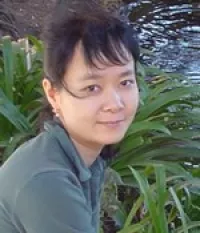
Rosemary Akhurst, PhD
Professor, Department of Anatomy
Director, Pre-clinical Therapeutics, Helen Diller Family Comprehensive Cancer Center
How do components of the TGF-b signaling pathway regulate mammalian developmental processes and disease outcomes in vivo? This is the overarching question addressed in the Akhurst lab. TGF-b is required for normal embryonic development, especially vascular development and immune tolerance. It is a major player in tumor progression, including epithelial mesenchymal transformation, immunosuppression, elaboration of extracellular matrix, cancer stem cell maintenance and drug resistance, all activities that drive tumor progression and metastasis. The laboratory studies pre- and post-natal vascular development, as well as mouse models of tumorigenesis. Our studies span basic cellular molecular biology and developmental genetics, through systems genetics, to translational studies on preclinical drug development, with translational validation using human genetics, bioinformatics and analysis of human tissues.
Christopher Allen, PhD
Associate Professor, Department of Anatomy, Cardiovascular Research Institute
My laboratory is interested in the cellular communication and differentiation programs in allergic immune responses, particularly in asthma. My laboratory is applying sophisticated imaging, flow cytometry, mouse genetics, and other techniques to uncover novel paradigms underlying allergic inflammation. As a major area of emphasis, we are studying the generation and function of IgE antibodies that initiate allergic inflammation. We generated fluorescent IgE reporter mice and allowing us to identify and track rare IgE-producing B cells in vivo. We also optimized a flow cytometry procedure to identify rare IgE-producing B cells in both mouse and human samples, facilitating mechanistic studies. Our work has revealed that the IgE B cell receptor exhibits antigen (ligand)-independent signaling that promotes plasma cell differentiation and limits IgE germinal center B cell responses. We are also imaging the lungs and associated lymphoid tissues by two-photon laser scanning microscopy to directly visualize cellular interactions in situ. Using this approach, we achieved the first in vivo analysis of the interactions of CD4 T cells with basophils, which are rare IgE effector cells. We are now analyzing the interactions of basophils with other cell types during secondary immune responses to further elucidate the functions of these cells. More broadly, we are considering the combined cellular interactions among various types of inflammatory cells in the lungs in mouse models of asthma.
Allan Basbaum, PhD
Professor, Chair, Department of Anatomy
Following upon our extensive earlier studies of the CNS circuits through which opioids exert their analgesic effects, our laboratory now examines the mechanisms through which tissue and nerve injury produce changes in the peripheral and central nervous system that result in persistent pain. In parallel studies, we examine the circuits through which pruritogens generate itch. The hallmark of our work is a multidisciplinary approach to the problem, using molecular, neuroanatomical, pharmacological and behavioral analyses in wild type, and genetically-modified mice, including knockouts and Cre- or reporter-expressing mice. By combining these studies with an analysis of the functional properties of molecularly-defined neurons, these studies examine the extent to which pain and itch circuits segregate or converge at the level of spinal cord interneurons and projection neurons. In recent studies, we turned our attention to the possibility of overcoming the neurological consequences of peripheral nerve damage, by transplanting embryonic cortical GABAergic precursor cells into the spinal cord. We have demonstrated that the cells integrate synaptically and functionally into host neural circuits and can ameliorate the persistent pain and itch associated with nerve damage. Very recently, we have significantly expanded the scope of our studies. We are using calcium imaging of cortical neurons in awake mice to examine the brain circuits and mechanisms through which pain and itch percepts are generated as well as the mechanisms through which general anesthetics exert their analgesic action.
Rohit Bose, MD, PhD
Assistant Professor, Department of Anatomy
“Unraveling transcription and evolution in hormone-related tumorigenesis”
Dr. Bose is a physician-scientist and practicing medical oncologist. Our group investigates hormone-related cancers, which possess unexploited vulnerabilities after developing hormone therapy resistance. We investigate how such tumors dynamically evolve through treatment cycles, and maintain compensatory transcriptional states. We use these findings to develop rational combinatorial therapies involving both existing and novel approaches.
Evan Feinberg, PhD
Assistant Professor, Department of Anatomy
Survival requires the flexibility to achieve one’s behavioral goals from virtually any starting point. This remarkable ability enables a hungry person to pick an apple regardless of whether their hand begins overhead or at their side, an imperiled mouse to escape to its burrow from anywhere in its environment, and a dragonfly to capture prey at any point in flight. We investigate how the brain flexibly selects movements that take into account the animal’s starting point and how this process is altered in conditions such as autism. Our multidisciplinary approach merges mouse behavior, neural recording and perturbation, anatomical tracing, molecular tool development, and mathematical modeling.
Leanne Jones, PhD
Professor, Department of Anatomy
Director, Bakar Aging Research Institute
In the Jones lab, our primary interest is in identifying conserved mechanisms that are involved in regulating stem cell behavior. To address such questions, my lab employs the Drosophila male germ line and intestine, as well as human intestinal tissue and 3D organoids, as model systems. We combine forward genetics, candidate approaches, genomics, and biochemistry to identify and characterize mechanisms that regulate the choice between stem cell self-renewal and initiation of differentiation, with a particular focus on the role of the local microenvironment, known as the “niche”. Once mechanisms that regulate stem cell behavior are identified, we evaluate how these mechanisms are altered byorganismal aging and chronic and acute changes in metabolism. Furthermore, we are particularly interested in understanding how dysregulation of stem cell behavior is linked to degenerative and/or age-onset diseases.
Christoph Kirst, PhD, MS
Assistant Professor, Department of Anatomy
Dr. Kirst studies how brains perform computations with a focus on how neuronal circuits achieve their flexible function and coordinate processing among different sub-networks.
He works in the interface between mathematics, physics, computer science, and neurobiology to build theoretical, mechanistic as well as conceptual understanding of flexible brain function. He collaborates closely with experimentalists on a broader range of model systems, data sets, and experimental paradigms to investigate the structure, dynamics, modulation, and function of single neurons, neuronal circuits, large-scale neuronal networks, and whole brains.
Carolyn Larabell, PhD
Professor and Vice Chair, Department of Anatomy
Mark Le Gros, PhD
Professor, Department of Anatomy
Our goal is the application of physics and engineering approaches to the study of cell behavior. In particular, we are developing a number of new imaging tools, such as soft x-ray tomograhy and cryogenic light microscopy, and establishing new methods for identifying and isolating cells displaying rare or dynamic phenotypes.
Qili Liu, PhD
Assistant Professor, Department of Anatomy
Animals can exhibit hunger for not only calories but also specific nutrients, which energize behaviors to search for and ingest particular types of food in response to internal nutritional deficiency. Liu lab aims to understand the neurobiological basis of nutrient-specific appetite, and investigate how does hunger for specific nutrients interact with each other to ensure that a balanced diet is consumed. To this end, the lab will harness the power of Drosophila model system and employ a multidisciplinary approach including large-scale genetic and behavioral analyses, immunohistochemistry, functional imaging, and patch-clamp electrophysiology. These studies will shed light on the fundamental principles for the organization and modulation of motivated behaviors. Given the global obesity epidemic, illuminating the regulation of food choice guided by nutrient specific appetite may facilitate the development of novel therapeutic treatments for obesity.
Donald McDonald, MD, PhD
Professor, Department of Anatomy
Research in the McDonald lab studies mechanisms and consequences of blood vessel changes in cancer and inflammation to determine the contributions of altered vascular growth and function to disease pathophysiology and treatment. In-vivo cell biological approaches have been developed for use in mouse models to learn how the vasculature remodels during disease progression and how these alterations can serve as therapeutic targets.
Ongoing experiments are examining the processes underlying defective endothelial barrier function in inflammation. New strategies are being explored to reverse vascular leakage by regulating signaling pathways that control the junctions between endothelial cells. Here the focus is the regulation of endothelial tight junctions and adherens junctions by angiopoietins, Tie1 and Tie2 tyrosine kinase receptors, and endothelial cell-specific phosphotyrosine phosphatases.
Other work is elucidating the antitumor actions of engineered oncolytic vaccinia viruses that home selectively to tumor blood vessels and reduce tumor growth. Viral infection of tumor blood vessels is followed by focal tumor infection accompanied by widespread tumor cell killing mediated by a robust immune response. Studies are examining immune checkpoint inhibitor amplification of the action of genetically modified vaccinia viruses on primary tumors and on metastases. The overall goal is to advance the mechanistic understanding of blood vessel growth and remodeling and to develop strategies that exploit changes in vascular phenotype in the treatment of cancer and inflammatory diseases.
Takashi Mikawa, PhD
Professor, Departments of Anatomy, Cardiovascular Research Institute
The establishment of extremely complicated structures and functions of our organ systems depends upon orchestrated differentiation and integration of multiple cell types. Our group focuses to explore a common developmental plan for successful organogenesis, by investigating the mechanisms involved in the differentiation and patterning of the cardiovascular and central nervous systems.
Keith Mostov, MD, PhD
Professor, Department of Anatomy
“The Mostov lab wants to understand how the shape, structure, and size of epithelial organs are determined during development, as well as how we can use that knowledge to foster the regeneration of damaged organs.”
Tomasz Nowakowski, PhD
Assistant Professor, Department of Anatomy
Dr. Nowakowski studies the principles of tissue development, the timing of cell generation, intercellular interactions, and developmental lineage relationships to uncover underlying neurodevelopmental events, tissue organization and cellular demographics.
Todd Nystul, PhD
Associate Professor, Department of Anatomy
Dr. Nystul’s laboratory uses the Drosophila ovary as a model for studying the fundamental properties of epithelial stem cells, their associated niche, and the connection between epithelial stem cells and cancer. They are interested in questions such as, (1) how is stem cell fate maintained within a dynamic epithelial tissue; (2) What is the nature of the epithelial stem cell niche; (3) What is the genetic basis of competition for the stem cell niche; and (4) Do stem cell defects contribute to cancer and other pathologies?
Rushika Perera, PhD
Assistant Professor, Department of Anatomy
Perera lab focuses on understanding how scavenging pathways such as autophagy and the lysosome - a degradative organelle - enables metabolic and cellular adaptation to stress and contributes to aggressive features of disease. Beyond mediating degradation of diverse macromolecules, the lysosome also plays an important role in signal transduction, cellular quality control, metabolism and detoxification. The lab has a particular interest in how the lysosome contributes to cancer pathogenesis with a current focus on studying pancreatic ductal adenocarcinoma (PDA). The lab prior studies have identified novel mechanisms mediating constitutive activation of the MiT/TFE family of transcription factors which regulate autophagy and lysosome biogenesis in PDA and in normal physiological contexts. Perera lab has also discovered new roles for autophagy and the lysosome in facilitating immune evasion and enhanced metabolic fitness of PDA cells. The lab ongoing research leverages their combined experience in cellular trafficking, autophagy and lysosome function combined with in vitro and in vivo model systems, organelle isolation and mass spectrometry-based proteomics to determine critical roles of the lysosome in cancer progression and metastasis, tumor heterogeneity, stress adaptation and drug resistance.
Jeroen Roose, PhD
Professor & Vice Chair, Department of Anatomy
The Roose lab focuses on understanding cell fate decisions driven by cell-cell interactions and signaling pathways, in the context of cancer, autoimmune diseases, and COVID-19. Our collaborative research programs benefit from many technology platforms, such as organoids, CyTOF, single cell RNAseq, spectral flow, mouse models, and patient samples. Dr. Roose is a co-founder of the Bakar ImmunoX initiative at UCSF https://immunox.ucsf.edu/ and a co-lead investigator on the UCSF Endeavor Program that focuses on cancer metastasis, funded through the Mark Foundation for Cancer Research
Visit the Roose Lab
Visit Roose Organoid disease to biology Plug-in CoLab
Saul Villeda, PhD
Assistant Professor, Department of Anatomy
Our lab is interested in understanding what drives regenerative and cognitive impairments in the aging brain, and moreover how the effects of aging can be reversed in the old brain. Our lab is focused on three areas. First, we are looking at how immune-related changes in old blood contribute to impairments in neural stem cell function and associated cognitive functions. Second, we are looking at the contribution of the innate immune system to age-related impairments in synaptic plasticity and cognitive function. Third, we are looking at how exposure to young and exercise blood rejuvenates neural stem cell function, synaptic plasticity and cognitive function in the old brain. Ultimately, our goal is to elucidate cellular and molecular mechanisms that promote brain rejuvenation as a means by which to combat age-related neurodegeneration and cognitive dysfunction.
Allison Xu, PhD
Professor, Department of Anatomy, Diabetes Center
Research in Allison Xu’s laboratory focuses on understanding the adaptive regulation of energy balance and glucose homeostasis in response to changes of physiologic and environmental conditions. Specifically, Allison’s lab is investigating how hypothalamic and hindbrain neurons sense and integrate peripheral hormones and nutritional signals, and how these neurons regulate feeding, energy expenditure, systemic glucose and lipid metabolism. These efforts will help better understand how energy balance and glucose homeostasis are regulated and how these regulations are disrupted in obesity, type 2 diabetes and aging.
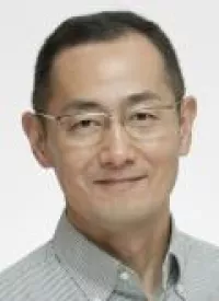
Shinya Yamanaka, MD, PhD
Professor, Department of Anatomy
Dr. Yamanaka’s research focuses on ways to generate cells resembling embryonic stem cells by reprogramming somatic cells. Over a decade ago, Dr. Yamanaka discovered that adult somatic cells can be reprogrammed into an embryonic-like pluripotent state by delivering transcription factors. These reprogrammed cells, known as induced pluripotent stem (iPS) cells, have the potential to develop into every cell type in the body and are invaluable tools for disease modeling, drug screening, and cell therapy. iPS cells also provide an unprecedented opportunity for discovery in life science, and in Yamanaka’s lab today, researchers continue to use them to investigate the mechanisms for cell fate determination, the reprogramming process, and pluripotency.
Primary Faculty | Affiliated Faculty | Teaching Faculty | Emeritus Faculty


